
External Gas Bubble Disease
Skin bubbles or cysts can be an indication of several different ailments and can appear as gas filled vesicles or appear solid or cystic in nature.
The clear, gas filled vesicles are usually diagnostic of External Gas Bubble Disease (EGBD).
Cystic lesions filled with pus are called abscesses are usually due to Vibrio infections although other bacterial infections are possible. Lesions that tend to be more solid in nature include granulomatous diseases such as Norcardia and Mycobacteria. Granulomata of the skin have also been seen in parasitic infections caused by nematodes. Biopsy of solid lesions, such as these, is the best route for definitive diagnosis. Besides parasites, granulomatous disease usually does not respond to therapy and euthanasia with system sterilization is recommended. Glugea is also a fatal, contagious non-treatable disease that presents with solid lesions that are not granulomatous. It is also easily diagnosed in biopsy material. - information provided by Renee Coles-Hix
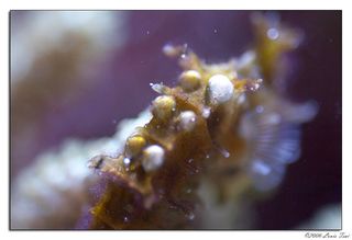 Hippocampus barbouri with gas bubbles
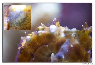 Hippocampus barbouri with gas bubbles
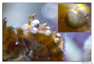 Hippocampus barbouri with gas bubbles
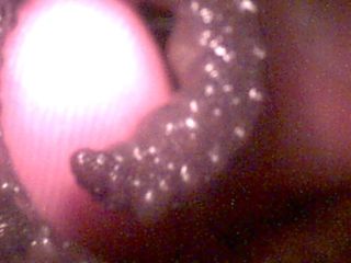 Gas Bubble on tail
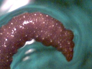 Gas Bubble on tail
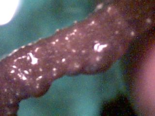 Gas Bubble on tail
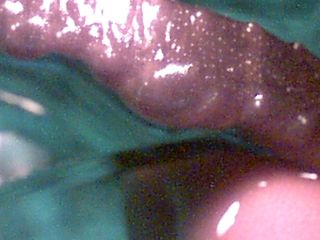 Gas Bubble on tail
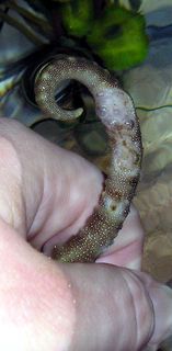 Bubbles on tail
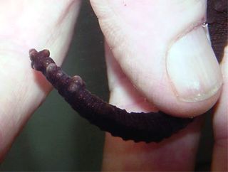 Gas bubbles in tail
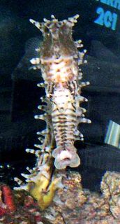 Hippocampus barbouri with snout bubbles
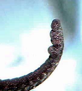 Tail with bubbles
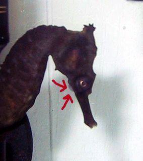 Trigger with bubbles underneath
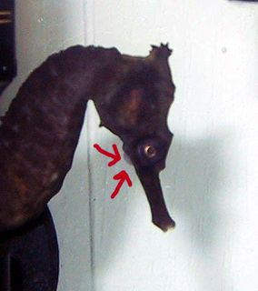 Trigger with bubbles underneath
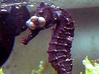 Severe gas bubbles surrounding the eyes
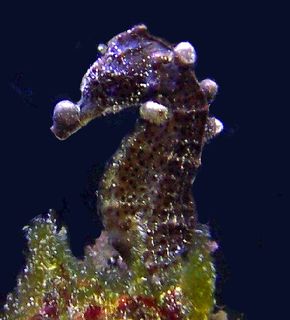 Kuda with gas bubbles on head and torso
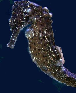 Kuda with gas bubbles on head and torso
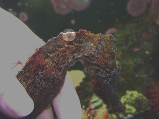 Female With Gas Bubble Disease
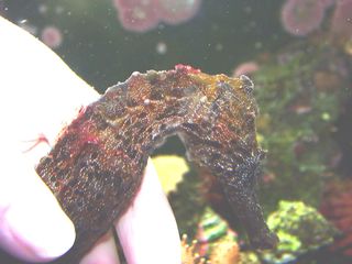 Female With Gas Bubble Disease
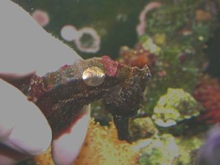 Female With Gas Bubble Disease
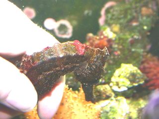 Female With Gas Bubble Disease
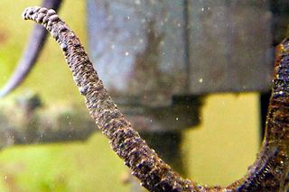 Tail with bubbles
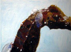 External Gas Bubble Disease Example
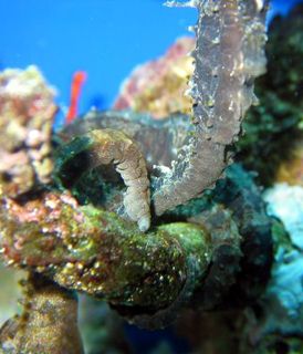 Gas bubbles on tail
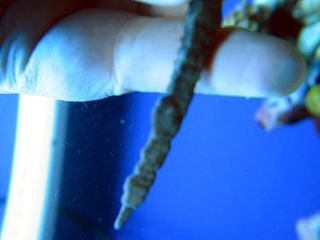 closeup of gas bubbles on tail
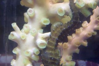 Large gas bubble on tail underside
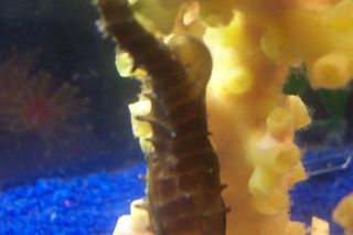 Large gas bubble on tail underside
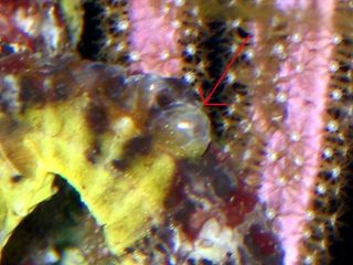 Large bubble on head
Photos are © their contributors and Seahorse.org. Please ask before reusing them.